What happens?
After you have been referred for an examination you should contact the practice and organise a booking. Be sure to ask:
- How much will it cost?
- How can I pay for the examination?
- How do I prepare for the examination?
Our medical reception staff are there to make your appointment process easy and are happy to answer all your questions.
Once your booking is made, simply turn up on the day and our specialist medical technicians will take you through the scheduled procedure. Once the procedure is completed a qualified Radiologist will interpret the images and issue a diagnostic report to your referring Doctor.
How long will it take?
Depending upon which examination you have, the procedure could take anything from 15mins to several days. The practice you book your appointment with will be able to confirm the length of time, as well as other important preparation information. You can check out approximate times and preparation details under the respective service listed above.
After Your Examination?
Most examinations allow you to leave immediately without restrictions. Check with the practice before you leave for any special instructions. Your results will either be sent to the referring Doctor or made available to you.
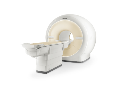
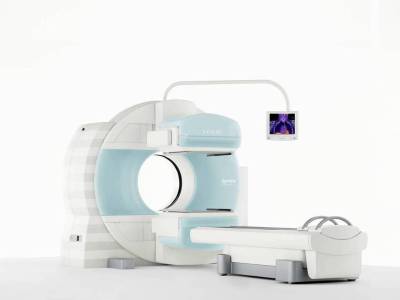
What is it?
Magnetic Resonance Imaging or MRI is and advanced diagnostic imaging technique that produces high-resolution, computerised images of the human body, sometimes in 3D. These images are photographed on film and reported by an MRI specialist radiologist before being sent to your referring doctor. MRI scanning does not use radiation and is a painless procedure.
Is MRI for everyone?
Unfortunately not everyone is able to have an MRI Scan. Some implants, including pacemakers and cochlear ear implants, exclude individuals from undergoing MRI.
What to tell us?
If you experience high levels of anxiety or claustrophobia or you have any metal or surgical implants.
If you have had metal in your eyes at anytime during your life and have not had a MRI scan before, an X-ray will be taken by our staff to ensure that no metal remains. Should metal be detected, it will need to be removed by a doctor prior to having a scan. Metal within your eyes may cause permanent eye damage to due to the high magnetic field of the MRI scanner.
How to Prepare?
No specific preparation is required. You may eat and drink normally and take your usual medication. If you are taking medication for pain, we advise that this is taken prior to the scan as it is important to remain still throughout the examination. Jewellery and makeup cannot be worn whilst having an MRI scan.
What does the procedure involve?
For the examination you will be asked to lay on a padded examination table that slowly glides into the scanner. You will be positioned comfortably, generally with your arms at your side.
The MRI scanner is well lit and remains open at both ends. Air consistently circulates around you during the examination. The scanner makes a series of loud banging noises and there will be a small amount of vibration while the images are being collected. The technicians will provide you with some ear plugs or headphones with music of your choice and they will be in constant contact with you during the test.
You must hold very still and may be asked to keep from breathing for a few seconds while the images are being taken to reduce the possibility of a blurred image.
How long will it take?
MRI procedures can take up to 1hr depending on the body part being scanned.

What is it?
CT stands for computerised tomography. CT is a diagnostic imaging technique that produces a sequence of detailed cross-sectional images of the body.
What must you tell us?
If you have had an adverse reaction to a previous contrast injection or other drugs or if you have any renal impairment.
If you take any diabetic medication that contains metformin (examples of which are diabex, diaformin, avandament, glucomet, novamet, genex, glucophage), insulin, or blood thinner (warfrin or aspirin products).
If you are pregnant or suspect you may be pregnant or breast feeding.
How to Prepare?
You should arrive 15 minutes before your appointment time to complete all necessary paperwork
You should wear comfortable clothing that have no metal zippers, belts or buttons in the abdominal or pelvic areas. You may be asked to remove jewellery, eyeglasses and metal objects that could interfere with the x-ray images. If you do not have clothing without metal components we may ask you to change into a gown.
Medications may be taken as prescribed.
You may be asked to stop eating solid foods 2 hours prior to the procedure.
Depending upon which part of the body is being scanned you may be required to drink an oral contrast 1 hour prior to your exam. If you are having scans of your abdomen and/or pelvis you will need to drink this. The contrast helps to outline the stomach and intestines so abnormalities can be identified.
Bring any previous x-ray and CT films with you to the appointment in case they are needed for comparison.
What does the procedure involve?
You will be taken into our procedure room and positioned on a CT table either head first or feet first, depending on the part of the body being scanned. Some CT scans require Intravenous (IV) injection of contrast medium into a vein, usually at your elbow or back of the hand. It helps to visualize veins and arteries and certain tissues, as well as your urinary tract.
If given IV contrast you may notice a hot flushed feeling, get a funny metallic taste or smell. It is also common to feel a sensation in the pelvis or bladder area which can make feel that you need to urinate or that you have urinated. This is just a sensation and you do not actually urinate.
These sensations are normal and only last 10-60 seconds
You move through the scanner and will be required to remain motionless for the length of the examination. Some studies will require you to hold your breath.
If you received a contrast injection, the IV cannula is removed from your arm before you go home.
After your procedure you will be able to resume all normal activities.
If you notice any pain, redness, and/or swelling at the IV site after you return home following your procedure, you should notify your physician.
How long will it take?
The CT procedure normally takes between 10 – 45 minutes.
What is it?
X-rays are widely used in general medicine for diagnostic imaging. X-rays produce images of the body, its organs, and other internal structures for diagnostic purposes. X-rays pass through body structures onto specially-treated plates and a “negative” type picture is made (the more solid a structure is, the whiter it appears on the film).
What to tell us?
If you are pregnant or suspect you may be pregnant. If you have had a recent barium x-ray procedure.
How to Prepare?
You should arrive 15 minutes before your appointment time to complete all necessary paperwork.
You should wear comfortable clothing. You may be asked to remove jewelry, eyeglasses and metal objects that could interfere with the x-ray images. If you do not have clothing without metal components we may ask you to change into a gown.
Bring any previous x-ray films with you to the appointment in case they are needed for comparison.
What does the procedure involve?
The type of procedure will determine your positioning, such as lying on a table, sitting, or standing, and the type of x-ray equipment used. You will be positioned on an x-ray table, that carefully places the part of the body that is to be x-rayed between the x-ray machine and a cassette containing the x-ray film.
You will have to hold your breath for a few seconds for x-rays of the chest and abdomen area but this is not necessary for x-rays of the arms, legs and head. Although you may be required to hold awkward positions for a short period of time, there is no discomfort from the x-ray exposure.
How long will it take?
The procedure will take 30 to 90 minutes.

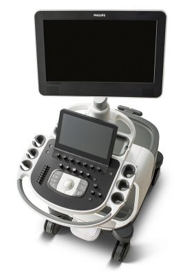
What is it?
Ultrasound is a procedure that uses sound waves to generate pictures of many parts of the body. Ultrasound can detect diseased or damaged tissues, locate abnormal growths and identify a wide variety of conditions. Pictures are created by applying gel to the skin and moving a transducer, (also known as an ultrasound probe), over the body part being examined.
Doppler Ultrasound
Doppler ultrasound uses high-frequency sound waves to obtain a picture of blood flow through various vessels in the body. It is commonly used to image the carotid artery in the neck, or arteries and veins in legs and arms. It can detect diseased vessels and identify a wide variety of changing conditions.
What to tell us?
If you have diabetes
How to Prepare?
You should arrive 15 minutes before your appointment time to complete all necessary paperwork.
Preparation will vary depending on the information your doctor needs and the type of exam you will have.
Wear comfortable clothing to your appointment. You may be asked to wear a gown for the procedure.
The preparations for an ultrasound vary depending on the body part you are having scanned.
If you are having an ultrasound of any Upper Abdominal Organ (Gall Bladder, Pancreas, Liver, Spleen) and/or Kidney(s) and/or Aorta eat an early low-fat dinner on the night before your exam and have nothing to eat or drink for 8 hours prior to your exam.
If you are having an ultrasound of your Pelvis you will need to have a full bladder for this exam. Please drink 1 litre of water one hour before the exam. Do not empty your bladder before the exam.
There is no preparation if you are having an ultrasound of your Breast, Extremity or other body parts (i.e., Thyroid).
Bring any previous x-ray films and ultrasounds with you to the appointment in case they are needed for comparison.
What does the procedure involve?
You may be asked to lie down on a table. Clothing over the area to be scanned will be removed. A special gel will be applied to the skin to prevent air from getting between your skin and the transducer. The transducer is then passed over the skin of the area being examined. The transducer generates and receives the high-frequency sound waves. The computer in the ultrasound unit processes and converts the resulting patterns into detailed images.
Normal activities can be resumed immediately after the test.
Doppler Vascular Ultrasound
A Doppler vascular ultrasound is performed the same way an Ultrasound is performed. During portions of the exam, you will hear sounds similar to a heartbeat coming from the ultrasound machine while we are listening to the blood flow in your vessels.
How long will it take?
Ultrasound procedures can take up to 60 minutes.
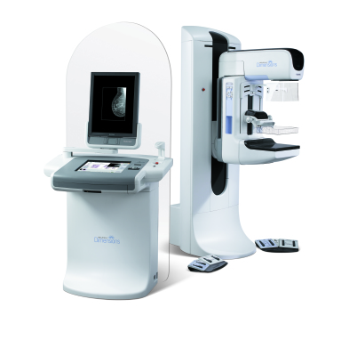
What is it?
Mammography is an X-ray examination of the breasts.
What to tell us?
If you have breast implants. If you are pregnant, suspect you may be pregnant, or are breastfeeding.
How to Prepare?
You should arrive 15 minutes before your appointment time to complete all necessary paperwork.
Wear comfortable clothing, including a two piece garment. Stop drinking caffeinated drinks several days before your mammogram appointment. Do not schedule your mammogram for the week before your period if your breasts are usually tender during this time.
If you take hormones (estrogen and progesterone), ask your doctor about the best time to schedule a mammogram.
Do not wear deodorant, powder, perfume or lotion on the day of your mammogram. These substances may obsure the images.
Remove all jewellery from your neck and chest area.
Bring any previous x-ray films, mammograms and breast ultrasounds with you to the appointment in case they are needed for comparison.
What does the procedure involve?
You’ll be asked to undress from the waist up and will be given a gown to wear. During mammography your breast is compressed between 2 plates attached to the mammogram machine. Compression is needed to keep your breast from moving, and to make it thinner. These measures reduce the x-ray exposure, reduce blurring, and make the image sharper. The x-ray pictures are taken from several angles. The process will be repeated for the other breast.
You must hold very still and may be asked to keep from breathing for a few seconds while the x-ray picture is taken to reduce the possibility of a blurred image.
How long will it take?
Mammogram procedures can take 20 to 30 minutes.
- Our machines:
- Detect 41% more invasive cancers (the ones that have spread outside of the milk duct into surrounding, healthy tissues).
- Reduce false-positives, decreasing recall rates by 15-40% - sparing women the anxiety of being called back for further testing.
- Better visualise masses, distortions and asymmetric densities. May reduce the number of unnecessary biopsies.
- Increase cancer detection in women with dense breasts (e.g. women in their 40s).
What is it?
Nuclear Medicine examinations involve the injection or ingestion of a small quantity of a radioactive material (radioisotope). The radioactive levels, in most cases are equal to or less than what you would receive from a routine x-ray or CT examination. The radioactive substance accumulates in the target area of interest which is then examined under a gamma camera, to produce a picture. Nuclear medicine examines function and structure, this means that it can show how an organ is working, not simply what it looks like and is commonly used for bone scans, thyroid studies, lung scans, cardiac stress tests, stomach, liver, kidney and gallbladder procedures.
What to tell us?
If you are pregnant or suspect you may be pregnant.
How to Prepare?
You should arrive 15 minutes before your appointment time to complete all necessary paperwork.
Preparation will depend on the type of examination you are having. The practice will provide this information at the time you make your appointment.
Bring any previous x-ray films with you to the appointment in case they are needed for comparison.
What does the procedure involve?
The radioisotope is administered by an IV injection in your arm. It may take a few minutes to days for the radioisotope to reach the specific area to be studied. If there is a long wait period, you will be free to leave the center and return for your scan several hours or days later.
You will be positioned on an examination table. You must remain as still as possible during imaging. Once in position, the radioisotope emits gamma rays that are detected by a special camera to produce images about the area of interest.
How long will it take?
Nuclear medicine procedures can take 30 minutes – several days